

NURS 6512 Week 8: Assessment of the Musculoskeletal System
Sample Answer for NURS 6512 Week 8: Assessment of the Musculoskeletal System Included After Question
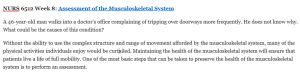
A 46-year-old man walks into a doctor’s office complaining of tripping over doorways more frequently. He does not know why. What could be the causes of this condition?
Without the ability to use the complex structure and range of movement afforded by the musculoskeletal system, many of the physical activities individuals enjoy would be curtailed. Maintaining the health of the musculoskeletal system will ensure that patients live a life of full mobility. One of the most basic steps that can be taken to preserve the health of the musculoskeletal system is to perform an assessment.
This week, you will explore how to assess the musculoskeletal system.
Learning Objectives
Students will:
- Evaluate abnormal musculoskeletal findings
- Apply concepts, theories, and principles relating to health assessment techniques and diagnoses for the musculoskeletal system
Photo Credit: SCIEPRO/Science Photo Library/Getty Images
Learning Resources
Note: To access this week’s required library resources, please click on the link to the Course Readings List, found in the Course Materials section of your Syllabus.
Required Readings
Ball, J. W., Dains, J. E., Flynn, J. A., Solomon, B. S., & Stewart, R. W. (2015). Seidel’s guide to physical examination (8th ed.). St. Louis, MO: Elsevier Mosby.
- Review of Chapter 4, “Vital Signs and Pain Assessment” (pp. 50-63)
- Chapter 21, “Musculoskeletal System” (pp. 501-543)
This chapter describes the process of assessing the musculoskeletal system. In addition, the authors explore the anatomy and physiology of the musculoskeletal system.
Dains, J. E., Baumann, L. C., & Scheibel, P. (2016). Advanced health assessment and clinical diagnosis in primary care (5th ed.). St. Louis, MO: Elsevier Mosby.
- Chapter 22, “Limb Pain” (pp. 356-374)
This chapter outlines how to take a focused history and perform a physical exam to determine the cause of limb pain. It includes a discussion of the most common tests used to assess musculoskeletal disorders.
- Chapter 24, “Low Back Pain (Acute)” (pp. 288-300)
The focus of this chapter is the identification of the causes of lower back pain. It includes suggested physical exams and potential diagnoses.
Sullivan, D. D. (2019). Guide to clinical documentation (3rd ed.). Philadelphia, PA: F. A. Davis.
- Chapter 2, “The Comprehensive History and Physical Exam” (“Muscle Strength Grading”; p. 29)
- Chapter 4, “Pediatric Preventative Care Visits” (“Documentation of Important Components of Age Specific Physical Exams and Sports Pediatric Sports Participation Physical Exam”; pp. 106-107)
Note: Download this Adult Examination Checklist and Physical Exam Summary: Abdomen to use during your practice musculoskeletal examination.
Katz, J. N., Lyons, N., Wolff, L. S., Silverman, J., Emrani, P., Holt, H. L., & …Losina, E. (2011). Medical decision-making among Hispanics and non-Hispanic Whites with chronic back and knee pain: A qualitative study. BMC Musculoskeletal Disorders, 12(1), 78–85.
Retrieved from the Walden Library databases.
This study examines the medical decision making among Hispanics and non-Hispanic whites. The authors also analyze the preferred information sources used for making decisions in these populations.
Smuck, M., Kao, M., Brar, N., Martinez-Ith, A., Choi, J., & Tomkins-Lane, C. C. (2014). Does physical activity influence the relationship between low back pain and obesity? The Spine Journal, 14(2), 209–216. doi:10.1016/j.spinee.2013.11.010
Retrieved from the Walden Library Databases.
Shiri, R., Solovieva, S., Husgafvel-Pursiainen, K., Telama, R., Yang, X., Viikari, J., Raitakari, O. T., & Viikari-Juntura, E. (2013). The role of obesity and physical activity in non-specific and radiating low back pain: The Young Finns study. Seminars in Arthritis & Rheumatism, 42(6), 640–650. doi:10.1016/j.semarthrit.2012.09.002
Retrieved from the Walden Library Databases.
Required Media
Online media for Seidel’s Guide to Physical Examination
Optional Resources
LeBlond, R. F., Brown, D. D., & DeGowin, R. L. (2014). DeGowin’s diagnostic examination (10th ed.). New York, NY: McGraw Hill Medical.
- Chapter 13, “The Spine, Pelvis, and Extremities” (pp. 585–682)
In this chapter, the authors explain the physiology of the spine, pelvis, and extremities. The chapter also describes how to examine the spine, pelvis, and extremities.
NURS 6512 Week 8: Assessment of the Musculoskeletal System
The body is constantly sending signals about its health. One of the most easily recognized signals is pain. Musculoskeletal conditions comprise one of the leading causes of severe long-term pain in patients. The musculoskeletal system is an elaborate system of interconnected levers that provide the body with support and mobility. Because of the interconnectedness of the musculoskeletal system, identifying the causes of pain can be challenging. Accurately interpreting the cause of musculoskeletal pain requires an assessment process informed by patient history and physical exams.
In this Discussion, you will consider case studies that describe abnormal findings in patients seen in a clinical setting.
Note: By Day 1 of this week, your Instructor will have assigned you to one of the following specific case studies for this Discussion. Also, your Discussion post should be in the Episodic/Focused SOAP Note format, rather than the traditional narrative style Discussion posting format. Refer to Chapter 2 of the Sullivan text and the Episodic/Focused SOAP Template in the Week 5 Learning Resources for guidance. Remember that all Episodic/Focused SOAP notes have specific data included in every patient case.
Case 1: Back Pain
A 42-year-old male reports pain in his lower back for the past month. The pain sometimes radiates to his left leg. In determining the cause of the back pain, based on your knowledge of anatomy, what nerve roots might be involved? How would you test for each of them? What other symptoms need to be explored? What are your differential diagnoses for acute low back pain? Consider the possible origins using the Agency for Healthcare Research and Quality (AHRQ) guidelines as a framework. What physical examination will you perform? What special maneuvers will you perform?
Case 2: Ankle Pain
A 46-year-old female reports pain in both of her ankles, but she is more concerned about her right ankle. She was playing soccer over the weekend and heard a “pop.” She is able to bear weight, but it is uncomfortable. In determining the cause of the ankle pain, based on your knowledge of anatomy, what foot structures are likely involved? What other symptoms need to be explored? What are your differential diagnoses for ankle pain? What physical examination will you perform? What special maneuvers will you perform? Should you apply the Ottowa ankle rules to determine if you need additional testing?
Case 3: Knee Pain
A 15-year-old male reports dull pain in both knees. Sometimes one or both knees click, and the patient describes a catching sensation under the patella. In determining the causes of the knee pain, what additional history do you need? What categories can you use to differentiate knee pain? What are your specific differential diagnoses for knee pain? What physical examination will you perform? What anatomic structures are you assessing as part of the physical examination? What special maneuvers will you perform?
To prepare:
With regard to the case study you were assigned:
- Review this week’s Learning Resources, and consider the insights they provide about the case study.
- Consider what history would be necessary to collect from the patient in the case study you were assigned.
- Consider what physical exams and diagnostic tests would be appropriate to gather more information about the patient’s condition. How would the results be used to make a diagnosis?
- Identify at least five possible conditions that may be considered in a differential diagnosis for the patient.
Note: Before you submit your initial post, replace the subject line (“Discussion – Week 8”) with “Review of Case Study ___.” Fill in the blank with the number of the case study you were assigned.
By Day 3
Post an episodic/focused note about the patient in the case study to which you were assigned using the episodic/focused note template provided in week 5 resources. Provide evidence from the literature to support diagnostic tests that would be appropriate for each case. List five different possible conditions for the patient’s differential diagnosis and justify why you selected each.
Note: For this Discussion, you are required to complete your initial post before you will be able to view and respond to your colleagues’ postings. Begin by clicking on the “Post to Discussion Question” link and then select “Create Thread” to complete your initial post. Remember, once you click on Submit, you cannot delete or edit your own posts, and you cannot post anonymously. Please check your post carefully before clicking on Submit!
Read a selection of your colleagues’ responses.
By Day 6
Respond to at least two of your colleagues on 2 different days who were assigned different case studies than you. Analyze the possible conditions from your colleagues’ differential diagnoses. Determine which of the conditions you would reject and why. Identify the most likely condition, and justify your reasoning.
Submission and Grading Information
Grading Criteria
Post by Day 3 and Respond by Day 6
To participate in this Discussion:
Assignment: Assessing the Skin, Hair, Nails, and HEENT
In preparation for the Head-to-Toe Physical Assessment Video due in Week 10, you will videotape yourself conducting an assessment of the skin, hair, nails, and HEENT this week.
To prepare:
- Arrange an appropriate time and setting with your volunteer “patient” to perform a skin, hair, nails, and HEENT examination.
- Submit your volunteer’s Video Release form prior to the exam.
- Download and review the Skin, Hair, and Nails and HEENT rubric provided in this week’s Learning Resources.
- Ensure that you have the appropriate lighting and equipment to perform the examination.
To complete:
- Record yourself performing the skin, hair, and nails, and HEENT physical examination. Be sure to cover all of the areas listed in the rubric and to use any equipment appropriately. This Assignment is due by Day 7 of Week 8. Submit your video using the Kaltura Mashup tool accessible through the Assignment submission link provided.
To submit your completed Video Assignment(s), do the following:
If you have not already done so, click on the Week 8 Assignment link. Once you have clicked on the link, click on the Write Submission button to turn on the Content Editor toolbar. Next, fill in the Submission field with any pertinent information. Attach your Assignment file by clicking on the Mashup button on the text editor menu bar and select Kaltura Media. Then find the media file you saved as “WK8Assgn+first initial+last name” and click on Open. Add any appropriate comments pertaining to your Assignment(s) in the Comments field. Be sure to attach all your video assignments. Finally, click on the Submit button to turn in your Assignment(s) for review.
For additional details on using the Kaltura Media mashup tool, please refer to the Kaltura Media Uploader page located in the course navigation menu.
By Day 7
This assignment is due.
Submission and Grading Information
Grading Criteria
Check Your Assignment Draft for Authenticity
To check your Assignment draft for authenticity:
Submit your Week 8 Assignment draft and review the originality report.
Submit Your Assignment by Day 7
Assignment 2 (Optional): Practice Assessment: Musculoskeletal Examination
A description of symptoms alone is not enough to form an accurate diagnosis of musculoskeletal conditions. Before forming a diagnosis, advanced practice nurses need to perform a physical examination. Although the musculoskeletal examination is relatively simple, it still needs to be performed multiple times before it can be mastered.
In preparation for the Head-to-Toe Physical Assessment Video due in Week 10, it is recommended that you practice performing a musculoskeletal examination this week.
Note: This is an optional practice physical assessment. You do not have to capture a video of this assessment, as no submission is required.
To prepare:
- Arrange an appropriate time and setting with your volunteer “patient” to perform a musculoskeletal examination.
- Download and review the Musculoskeletal Checklist provided in this week’s Learning Resources.
To complete:
- Perform the musculoskeletal examination. Be sure to cover all of the areas listed in the checklist.
A Sample Answer For the Assignment: NURS 6512 Week 8: Assessment of the Musculoskeletal System
Title: NURS 6512 Week 8: Assessment of the Musculoskeletal System
Patient Information:
A.J., 42, Male, Caucasian
CC: Back pain.
HPI: A.J. is a 42-year-old Caucasian male who presents to the clinic with complaints of lower back pain. He has been experiencing this pain for the past one month. Onset was initially abrupt and mild and has gradually worsened. He describes the pain a 4/10 most of the time. The pain is describes as a burning pain which radiates to the left leg at times. He experiences occasional numbness to the left leg and occasional sharp pains that he would rate 7/10. The pain is exacerbated with movement and relieved with rest and sitting/reclined.
Current Medications:
1.) Ibuprofen 400mg PO q6 hrs PRN for pain
2.) Multivitamin PO once daily in am
3.) Omeprazole 40mg PO once daily in am before breakfast
Allergies:
PCN – hives
Sulfa – angioedema
PMH
1.) Overweight
2.) Gastroesophageal reflux (GERD) – controlled
3.) Tobacco use
PSH
1.) Vasectomy 2014
2.) Cholecystectomy 2010
Sexual/Reproductive History:
Heterosexual
Vasectomy 2014
Social Hx:
Negative for current or past illicit drug use. Smokes 1 pack of cigarettes per day. Drinks alcohol in moderation.
Immunization History
Tetanus: 2014
Influenza: 11/2018
Family Hx:
Mother, alive, age 67, hx HTN, hypothyroidism
Father, alive, age 70, hx DM, HTN
Maternal Grandmother, alive, age 89, osteoporosis, dementia, CHF
Maternal Grandfather, deceased at age 82, MI, hx HTN
Paternal Grandmother, deceased at age 87, breast cancer, hx hypothyroidism
Paternal Grandfather, deceased at age 88, kidney failure, hx CKD, DM
Brother, alive, 45, depression
Sister, alive, age 38, GERD
Daughter, alive, 13, healthy, no known medical issues
Lifestyle:
Patient is a self-employed farmer, married for 16 years with 1 dependent child. Has support of wife and daughter. Diet is high in meat and carbohydrates, consisting of three meals a day, mostly home cooked. Claims he does not regularly exercise but does have a physically demanding and quite stressful job. Has health insurance through his wife’s employer. Has not seen his primary care provider within the past year.
ROS:
General: No weakness or night sweats. No recent weight gain or loss of significance. Denies recent illness, fever, chills, or feeling fatigued.
HEENT: No history of head injury. No corrective lenses. Denies visual changes, diplopia, floaters, or photophobia. Denies any hearing difficulties or loss of hearing. Denies tinnitus, vertigo, or infections. Occasional sinus drainage, seasonal. Denies any change in sense of smell. Denies any episodes of epistaxis, nasal polyps, or recent sinus infection. Denies bleeding gums; cavities that have been filled. Reports good oral care, last dental visit was 6 months ago. Denies difficulty chewing or swallowing.
Neck: Denies lumps, swollen glands, pain, or stiffness.
Breasts: Denies lumps or pain.
Respiratory: Denies cough, shortness of breath, or night sweats.
Cardiovascular: Denies chest pain, pressure, palpitations, or orthopnea.
Gastrointestinal: No recent nausea, vomiting, diarrhea, or constipation. GERD controlled. No melena or hematochezia. No pain, appetite is good. No known liver problems, gallbladder removed 2010.
Genitourinary: No frequency, urgency, dysuria or hematuria. Denies incontinence or loss of bowel or bladder function, pain, or discharge. No prostate exam to date. No known hernia, masses.
Peripheral vascular: Denies varicose veins or edema. Experiencing low back pain which radiates to left leg, burning pain with occasional numbness and sharp pain to left leg. Capillary refill < 3 seconds.
Musculoskeletal: Low back pain x1 month; radiates to left leg at times. Occasional sharp pain and numbness to left leg. Denies recent fall or trauma.
Integumentary: No rash or itching. Denies dermatitis or psoriasis.
Psychiatric: Denies history of psychiatric disorders. No thoughts of self-harm. Denies depression and anxiety.
Neurologic: Denies headache, weakness, seizures, syncope, stumbling, or changes in balance or coordination. Denies changes in speech or memory. Admits to occasional numbness to left leg.
Hematologic: Denies anemia, bleeding, bruising, or history of clotting disorders. No history of blood transfusion.
Endocrine: No night sweats, cold or heat intolerance, polyuria. No excessive thirst or hunger.
Allergic/Immunologic: Denies asthma, eczema, or rhinitis. No known immune deficiencies.
Physical Examination
Vital signs:
BP: 136/76, right arm, sitting; HR: 91, regular; RR: 16, regular; T: 98.2 degrees F, typanic; SpO2: 97% RA; W: 210 pounds, stable; Ht: 5’7”; BMI: 32.9
General Appearance: Alert and oriented x3, cooperative, and answers appropriately. Patient appears to be in discomfort but in no acute distress. Well-groomed, appropriately dressed, overweight.
HEENT: Hair of average texture. Scalp without lesions, normocephalic/atraumatic. Conjunctiva pink; sclera white. Pupils equal, round, regular, reactive to light. Extraocular movements intact. Tympanic membranes visualized, clear canal and good cone of light, bilaterally. Acuity good to whispered voice. Mucosa pink, septum midline. Oral mucosa pink. Good dentition. Tongue midline, pink, and moist. Tonsils absent. Pharynx without exudates.
Neck: Trachea midline, supple, no palpable nodes
Lymph nodes: No lymphadenopathy in any nodes. No palpable cervical, axillary or epitrochlear nodes. Small inguinal nodes bilaterally, soft and nontender.
Chest: Heart rate regular with normal S1, S2; no S3, S4. No murmurs, rubs, and gallops.
Lungs: Lung expansion symmetrical, regular and non-labored, CTA without rales, wheezes or rhonchi.
Peripheral vascular: No pedal edema; 2+ dorsalis pedis pulses bilaterally, capillary refill less than 3 seconds.
Musculoskeletal: No obvious deformities, masses, discoloration, or enlarged joints. Palpable pain to lower lumbar region. No palpable spasms. ROM limited; able to bend side to side but with difficulty when twisting and going into extension. SLRs were positive. CLRs were negative.
Neurologic Mental Status: Awake, alert and oriented to person, place, and time. Cooperative. No slurred speech. Deep tendon reflexes 2+lower extremity. SLR positive, sensory neurology intact to light touch. Patient able to toe and heel walk. Gait was stable. No limping noted.
Skin: Warm, moist, pale. Intact without lesions, rashes, or urticaria.
Diagnostics:
1.) X-ray – identify potential tumor or structural abnormality
2.) CT – rule out disc disease or tumor
3.) MRI – rule out disc disease or tumor
4.) Labs – CBC, ESR, serum calcium, alkaline phosphatase
(Hollier & Hensley, 2011).
5.) Urinalysis – assess kidney function – rule out pyelonephritis
(Dains, Bauman, & Scheibel, 2016).
Sciatic stretch test – Patient lies in supine position, elevates affected leg in attempt to provoke pain. This test would assess for sciatica (Hollier & Hensley, 2011).
Assess deep tendon reflexes and range of motion. These exams would assess for location of injury or disorder (Dains, Bauman, & Scheibel, 2016).
Assessment:
DDx:
When considering differential diagnoses of acute low back pain, it is important to consider possible origins as outlined by the Agency for Healthcare Research and Quality (AHRQ) guidelines. These guidelines help providers recognize signs and symptoms of conditions that are potentially serious, first such as a spinal fracture, infection, or tumor. The AHRQ guidelines then help the provider with other causes such as sciatica, back problems that are nonspecific, abdominally involved, or psychologically involved (Dains, Bauman, & Scheibel, 2016).
1. Sciatica (primary dx)
- Low back pain
- Burning sensation
- Radiates to leg
- Numbness along dermatomal area
- SLR produces pain
- Sitting knee extension produces pain
- No change sin bowel or bladder function
(Dains, Bauman, & Scheibel, 2016).
2. Lumbar strain
- Pain located in lumbar region
- Pain can radiate
- No changes in bowel or bladder function
(Rhoads & Jensen, 2015).
- Pain relieved with sitting
(Dains, Bauman, & Scheibel, 2016).
3. Herniated disc
- Pain located in lumbar region
- Pain can radiate
- Positive straight leg raise
(Rhoads & Jensen, 2015).
4. Spinal fracture
- History of strenuous lifting
- Pain felt at site of injury
(Dains, Bauman, & Scheibel, 2016).
5. Spondylolisthesis
- Pain occurs between L5 and S1
- Forward flexion may be limited
(Dains, Bauman, & Scheibel, 2016).
- Pain may radiate down leg
- May feel numbness in affected extremity
(Cleveland Clinic, 2014).
References
Cleveland Clinic. (2014). Spondylolisthesis. Retrieved from
https://my.clevelandclinic.org/health/diseases/10302-spondylolisthesis
Dains, J., Bauman, L., Scheibel, P. (2016). Advanced health assessment and clinical
diagnosis in primary care (5th ed.). St. Louis: Missouri: Elsevier.
Hollier, A., & Hensley, R. (2011). Clinical Guidelines in Primary Care: A Reference and
Review Book. Layfayette, LA: Advanced Practice Education Associated, Inc.
Rhoads, J. & Jensen, M. (2015). Cough. In J. Rhoads & M. Jensen (Eds.). Differential
diagnosis for the advanced practice nurse. (pp. 37-47). New York, NY: Springer Publishing Company.
A Sample Answer 2 For the Assignment: NURS 6512 Week 8: Assessment of the Musculoskeletal System
Title: NURS 6512 Week 8: Assessment of the Musculoskeletal System
Patient Information:
N.S., 42, M, AA
CC (chief complaint)” lower back pain.”
HPI: 42-year old AA male with a chief complaint of lower back pain for the past month. He states that at times this pain may radiate to his left leg. He describes the pain as being sharp and sometimes a burning sensation that goes down his left leg. Ibuprofen makes the pain tolerable. Walking and sitting for a long period of time makes it worst. The pain is 9/10 on a pain scale.
Current Medications: Ibuprofen 200mg x 2 tablets every 4 hours for pain. Multivitamin 1 tablet daily.
Allergies: bananas (itchy throat)
PMHx: all immunization up-to-date. Last tetanus shot 2017. Broke arm at age 7-year old from football.; wore a cast for 2 months. Denies any recent falls. Denies head or neck injuries.
Soc Hx: High school math teacher x 16 years. Enjoys fishing and camping Deny smoking and recreational drug use and alcohol consumption. Wears a seatbelt majority of the time while in a motor vehicle. Lives alone in a single-family home in a gated community. Recently divorced x 6 months. Has one daughter age 10.
Fam Hx: Both parents are living. Father is a police officer, has gout. Mother is a principal at a local high school; healthy and exercises 3x week. Paternal grandparents are living, healthy, and are military veterans. Maternal grandparents healthy, retired and lives in the surrounding neighborhood. Has one sister that is in the Army, healthy.
ROS:
GENERAL: No weight loss, fever, chills, weakness or fatigue.
HEENT: Eyes: wears glasses for astigmatism. No visual loss, blurred vision, double vision or yellow sclerae. Ears, Nose, Throat: No hearing loss, sneezing, congestion, runny nose or a sore throat.
SKIN: No rash or itching. Has the letters “N” and “S” tattooed on bilateral triceps.
CARDIOVASCULAR: No chest pain, chest pressure or chest discomfort. No palpitations or edema.
RESPIRATORY: No shortness of breath, cough or sputum.
GASTROINTESTINAL: No anorexia, nausea, vomiting or diarrhea. No abdominal pain or blood.
GENITOURINARY: NO burning on urination.
NEUROLOGICAL: No headache, dizziness, syncope, positive for burning sensation in the left leg. No change in bowel or bladder control.
MUSCULOSKELETAL: positive for lower back pain.
HEMATOLOGIC: No anemia, bleeding or bruising.
LYMPHATICS: No enlarged nodes. No history of splenectomy.
PSYCHIATRIC: No history of depression or anxiety.
ENDOCRINOLOGIC: No reports of sweating, cold or heat intolerance. No polyuria or polydipsia.
ALLERGIES: bananas. gets an itchy throat
Physical exam: General: Head and Neck: able to move the head up and down; side to side without any pain or difficulty. Back: Upper back non-tender to touch. Skin is smooth. Spine appears straight. No abnormal curves. Lower back tender to touch on the left side. No nodules noted. Unable to raise left leg greater than 70 degrees, while lying in a supine position (nerve root L5 or S1). Unable to squat without grimacing. Unable to extend left knee while sitting. Facial grimaces noted. Observed walking with a cane.
Diagnostic results: X-ray of the lumbosacral spine may reveal a bone spur, which is an overgrowth of bone that can be pressing on a nerve (Mayo Clinic, 1998-2019). MRI may show a herniated disk and/or inflamed soft tissue. Straight leg test, which can detect tension on the L5 and/or S1 nerve root (Agency for Health Care Policy and Research, 1992-1996). Dorsiflexion the left ankle can aggravate the L5 or S1 nerve root, which may be related to disc herniation (Agency for Health Care Policy and Research, 1992-1996). Sitting knee extension, which can be an indication of sciatica nerve root damage (Agency for Health Care Policy and Research, 1992-1996). A bone scan to rule out spine tumors (Agency for Health Care Policy and Research, 1992-1996). Labs: CBC, UA, and ESR to rule out infection.
- A.
Differential Diagnoses
- Herniated disk: most common cause of lower back pain that puts pressure on the spinal cord and nearby nerve roots (OrthoInfo , 1995-2018). When the nerve root becomes irritated, it causes pain, numbness, and weakness in one or both of your legs (OrthoInfo , 1995-2018).
- Radiculopathy/sciatica: Is often unilateral and secondary to compression or inflammation of a spinal nerve (Yishay, 2012). Pain can be reproduced during certain activities, such as sitting and walking (Yishay, 2012).
- Spinal stenosis: a narrowing of the spaces within your spine, which can put pressure on the nerves that travel through the spine that occurs most often in the lower back (Mayo Clinic, 1998-2019). May or may not have any symptoms. The symptoms may include pain, tingling, numbness and muscle weakness (Mayo Clinic, 1998-2019).
- Nephrolithiasis: the presence of crystalline stones (calculi) within the urinary system.
- Spinal neoplasia: Maybe suspected if lower back pain does not get better between 4-6 weeks after conservative treatments (Epocrates, 2019).
P.
This section is not required for the assignments in this course (NURS 6512) but will be required for future courses.
References
References
Agency for Health Care Policy and Research. (1992-1996). AHCPR Quick Reference Guides. Retrieved from Agency for Health Care Policy and Research (: https://www.ncbi.nlm.nih.gov/books/NBK52120/#_A34341_.
Epocrates. (2019). Musculoskeletal lower back pain. Retrieved from Epocrates: https://online.epocrates.com/u/2935778/Musculoskeletal+lower+back+pain/Diagnosis/Differential
Mayo Clinic. (1998-2019). Sciatica. Retrieved from Mayo Clinic: https://www.mayoclinic.org/diseases-conditions/sciatica/diagnosis-treatment/drc-20377441.
Mayo Clinic. (1998-2019). Spinal stenosis. Retrieved from Mayo Clinic: https://www.mayoclinic.org/diseases-conditions/spinal-stenosis/symptoms-causes/syc-20352961.
OrthoInfo . (1995-2018). Herniated Disk in the Lower Back. Retrieved from American Academy of Orthopedic Surgeons: https://orthoinfo.aaos.org/en/diseases–conditions/herniated-disk-in-the-lower-back/.
Yishay, A. B. (2012, April 25). Lumbar Radiculopathy. Retrieved from Spine Health: https://www.spine-health.com/conditions/lower-back-pain/lumbar-radiculopathy.
A Sample Answer 3 For the Assignment: NURS 6512 Week 8: Assessment of the Musculoskeletal System
Title: NURS 6512 Week 8: Assessment of the Musculoskeletal System
Patient Information:
Initials: A.M Age: 42 Sex: Male Race: Caucasian
CC (chief complaint): Lower back pain
HPI: 42 year old Caucasian male complaining of pain in his lower back for the last month. Patient states the pain also radiates to his left leg.
Location: Lower back
Onset: 1 month ago
Character: aching, turns into burning as day goes on
Associated signs and symptoms: Pain radiating to left leg with burning
Timing: comes and goes, worse upon waking up
Exacerbating/ relieving factors: laying down relives the pain but walking and standing up makes it worse
Severity: 5/10
Current Medications: Denies current medications other than OTC multivitamin daily and Motrin as needed for the pain
Allergies: Allergic to bananas
PMHx: Up to date on all immunizations. Tetanus shot 10/2018, flu shot also 10/2018
Soc Hx: Works as a delivery driver for UPS. Likes to fish and golf when not working. Married for 20 years with 2 kids. Denies tobacco use. ETOH occasionally on weekends. Always wears seatbelt while in the car. Good support system with wife and kids.
Fam Hx: Mother: Hx of cataracts, Father denies medical history, Children both have mild asthma.
ROS: cover all body systems that may help you include or rule out a differential diagnosis You should list each system as follows: General: Head: EENT: etc. You should list these in bullet format and document the systems in order from head to toe.
Example of Complete ROS:
GENERAL: No weight loss, fever, or fatigue.
HEENT: Eyes: No visual loss or blurred vision. Last eye exam 09/2018. Ears, Nose, Throat: No hearing loss, sneezing, sore throat, or trouble swallowing.
SKIN: No discoloration or rash.
CARDIOVASCULAR: No chest pain, chest pressure or chest discomfort.
RESPIRATORY: No shortness of breath or cough.
GASTROINTESTINAL: No nausea, vomiting or diarrhea.
GENITOURINARY: No burning or frequency with urination.
NEUROLOGICAL: No headache, dizziness, syncope, paralysis, ataxia, numbness or tingling in the extremities. No change in bowel or bladder control. No headaches.
MUSCULOSKELETAL: Lower back pain that radiates down left leg.
HEMATOLOGIC: No bleeding or bruising.
LYMPHATICS: No enlarged nodes. No history of splenectomy.
PSYCHIATRIC: No history of depression or anxiety.
ENDOCRINOLOGIC: No reports of sweating, cold or heat intolerance.
ALLERGIES: Allergic to Bananas. No medication or seasonal allergies.
Physical exam: Alert and oriented x3, BP 126/80, HR 84, RR 18, Temp 98.4, O2 sat 100%. Ht 6’3, Wt 225.
Musculoskeletal: Full ROM in all joints. BUE and BLE symmetrical. Extremities symmetrical in length, alignment, and position.
Diagnostic results:
X-ray of spine to show any fracture or damage
CT of spine to show any fractures, infections, or tumors
MRI to determine if there are any herniated disks
- A.
Differential Diagnoses:
Lower back strain- Most common cause of lower back pain. Occurs after an event and produces immediate lower back pain. Symptoms include lower back pain that radiates and muscle spasms.
Osteoarthritis- Deterioration in cartilage covering the synovial joints. Pain usually occurs in lower back. Symptoms include pain, numbness, and tingling.
Lumbar stenosis- narrowing of the spinal canal. This causes pain with walking or standing. Pain relief comes from sitting or bending forward.
Herniated disk- usually occurs in those ages 30-50 and will cause postural changes.
Spinal compression fracture- caused by a loss of bone mass that occurs with aging. Symptoms include back pain, loss of height, and hunched-forward posture.
References
https://www.webmd.com/osteoporosis/guide/spinal-compression-fractures-causes
Ball, J. W., Dains, J. E., Flynn, J. A., Solomon, B. S., & Stewart, R. W. (2015). Seidel’s guide to physical examination (8th ed.). St. Louis, MO: Elsevier Mosby.
Dains, J. E., Baumann, L. C., & Scheibel, P. (2016). Advanced health assessment and clinical diagnosis in primary care (5th ed.). St. Louis, MO: Elsevier Mosby.
A Sample Answer 4 For the Assignment: NURS 6512 Week 8: Assessment of the Musculoskeletal System
Title: NURS 6512 Week 8: Assessment of the Musculoskeletal System
Patient Information:
JR, 15y/o, male, Caucasian
A 15-year-old male reports dull pain in both knees. Sometimes one or both knees click, and the patient describes a catching sensation under the patella.
CC “Sometimes I have pain to one or both knees, and it sounds like a click”
HPI: This is a 15y/o active Caucasian male that presents with a c/o knee pain in which onset was about 3 days ago when he first noticed it after the big soccer game. Pt states the pain is dull and he can sometimes auscultate a clicking sound when he moves. The patient denies any n/v, fever or rash, but does notice some swelling in both knees with a catching sensation behind the patella. JR states the pain is worse upon strenuous activity, such as sports after school and going down flights of stairs. Pt. states he uses ice to relieve the pain and reduce swelling. Jr describes the severity of the pain as a 3/10 when at rest and a 7/10 when at play.
Current Medications: NONE
Allergies: NKA
PMHx: Pt up to date on all childhood immunizations including HPV vaccine taken 10/26/2018. Pt h/o childhood asthma, and chicken pox. No known surgeries or major illnesses, or hospitalizations noted.
Soc Hx: JR is a straight ‘A’ student, is active in school, plays soccer and basketball, and does not drink alcohol or smoke. JR, however, admits that he has a girlfriend and does practices safe sex, utilizing birth control and condoms. Pt has a healthy relationship with his parents and has 2 older siblings that are also active in sports and have had similar knee pains w/ surgical repair.
Fam Hx: Pts mother and father are both living and healthy as well as both maternal grandparents. Pt’s paternal grandfather died of lung cancer at age 65 years, and paternal grandmother had rheumatoid arthritis and died of ovarian cancer at age 53 years.
ROS:
GENERAL: No weight loss, fever, chills, weakness or fatigue.
HEENT: Eyes: No visual loss, blurred vision, double vision or yellow sclerae. Ears, Nose, Throat: No hearing loss, sneezing, congestion, runny nose or sore throat.
SKIN: No rash or itching.
CARDIOVASCULAR: No chest pain, chest pressure or chest discomfort. No palpitations.
RESPIRATORY: No shortness of breath, cough or sputum.
GASTROINTESTINAL: No anorexia, nausea, vomiting or diarrhea. No abdominal pain or bloody stools.
GENITOURINARY: No burning on urination
NEUROLOGICAL: No headache, dizziness, syncope, paralysis, ataxia, numbness or tingling in the extremities. No change in bowel or bladder pattern
MUSCULOSKELETAL: Swelling noted to bilateral knees w/ painful and limited mobility with movement
HEMATOLOGIC: No anemia, bleeding or bruising.
LYMPHATICS: No enlarged nodes. No history of splenectomy.
PSYCHIATRIC: No history of depression or anxiety.
ENDOCRINOLOGIC: No reports of sweating, cold or heat intolerance. No polyuria or polydipsia.
ALLERGIES: History of asthma, no hives, eczema or rhinitis.
VS: B/P 110/75: P 65; R 16; T 98.0; O2 100%; Wt 189lbs.; Ht 70.”
Physical exam:
General: Pt alert/oriented and appears to be a healthy active young man.
Head: Normocephalic
EENT: PEERL, EOMI, OP benign, no LAD
Respiratory: Lungs clr. to auscultation bilaterally. Non-labored with no wheezing noted
Cardiovascular: Regular rate, rhythm, no murmur noted. A cardiac examination on male athletes is very important and should include auscultation with provocation maneuvers to screen for hypertrophic cardiomyopathy which is the most common cause of sudden death in this age group (Sullivan, 2019).
Musculoskeletal: Swelling noted to bilateral knees, w/ popping sound ascultated upon manipulation of bilateral knees using the muscle strength test (MST). MST tests for lower extremities extensor and flexor abilities against resistance against both distal and proximal groups. Knees warn to touch.
Integumentary: Skin intact, no punctures, rashes, abrasions, redness
Neurologic: All pulses palpable in all extremities, w/o focal deficit
Diagnostic results: Complete blood count (CBC) to r/o infection or leukemia associated with neoplasm (Dains, Baumann, & Scheibel, 2016). Erythrocyte Sedimentation Rate (ESR) a test for the non-specific measurement of inflammation (Dains, Baumann, & Scheibel, 2016). Antinuclear antibody (ANA) are positive in the case of Rheumatoid arthritis (RA) and Systemic Lupus Erythematosus (SLE) (Dains, Baumann, & Scheibel, 2016). Rheumatoid factor (RF) to confirm the diagnosis of rheumatoid arthritis (2016). RF can be present for years without being symptomatic (2016). 2View bilateral knee Xray to rule out fracture or dislocation (2016). Computed Tomography (CT) for further visualizations of bones in knee followed by a Magnetic resonance imaging (MRI) for possible soft tissue injuries (2016).
- A.
Presumptive Diagnosis: Meniscal Tear
Differential Diagnoses :
1. Meniscal Tear: will cause the inability for patients to straighten or bend the knee but the movement is usually painful (Dains, Baumann, & Scheibel, 2016). A sudden twisting injury is probably related to a meniscal tear and serious ligament disruption (Dains, Baumann, & Scheibel, 2016). When the knee locks abruptly and the patient complains of something getting in the way with the inability to extend the knee fully is a sign of a chronic unstable meniscus tear (2016).
2. Anterior Cruciate Ligament (ACL) Tear: With an ACL tear a loud pop is indicative of an ACL tear (Dains, Baumann, & Scheibel, 2016). Pediatric ACL tears are very rare and account for <5% of all ACL injuries and rarely occur in children under the age of 9 (Siebold, Seil, & Engebretsen, 2015). Because this is considered a serious injury of the knee, studies found associated injuries in kids and adolescents as high as 50-65% (Siebold, Seil, & Engebretsen, 2015).
3. Stress Fracture: When adolescents are unable to tolerate intense training trabecular microfractures develop in the bone resulting in bone breakage (Dains, Baumann, & Scheibel, 2016). Healthy children who participate in high-impact physical activity have been known to improve bone health during growth and development (Tenforde, Lynn Sainani, Carter Sayres, Milgrom, & Fredericson, 2014). However, a subset of athletes may be at an increased risk for stress fractures, with females at a greater risk than males (Tenforde, Lynn Sainani, Carter Sayres, Milgrom, & Fredericson, 2014).
4. Chondromalacia Patellae (CP): is most common in adolescent females, but forms as a change in the in the patellofemoral joint cartilage (Dains, Baumann, & Scheibel, 2016). The onset is caused by trauma, misalignment of the patella, and anatomic anomalies (Dains, Baumann, & Scheibel, 2016).
5. Patellar Tendinitis (PT) (jumper’s knees): this condition is characterized by inflammation of the distal extensors of the knee joint (Dains, Baumann, & Scheibel, 2016). PT is more common in athletes who place extra strain on knees from running and jumping (Dains, Baumann, & Scheibel, 2016). Signs are usually distinguished by complaint of dull, achy knee pain along with a clicking or popping sound (Dains, Baumann, & Scheibel, 2016).
P.
N/A
References
Dains, J. E., Baumann, L. C., & Scheibel, P. (2016). Advanced health assessment and
clinical diagnosis in primary care (5th ed.). St Louis, MO: Elsevier Mosby.
Siebold, R., Seil, R., & Engebretsen, L. (2015). ACL tear in kids: serious injury with high risk of
osteoarthritis. Knee Surgery, Sports Traumatology, Arthroscopy, 24(3), 641-643.
doi:10.1007/s00167-015-3912-1
Sulliavn, D.D. (2019). Giude to clinical documentation (3rd ed.). Philadelphia, PA: F. A.
Davis.
Tenforde, A. S., Lynn Sainani, K., Carter Sayres, L., Milgrom, C., & Fredericson, M. (2014).
Participation in Ball Sports May Represent a Prehabilitation Strategy to Prevent Future
Stress Fractures and Promote Bone Health in Young Athletes. PM&R, 7(2), 222-225.
doi:10.1016/j.pmrj.2014.09.017
A Sample Answer 5 For the Assignment: NURS 6512 Week 8: Assessment of the Musculoskeletal System
Title: NURS 6512 Week 8: Assessment of the Musculoskeletal System
A 46-year-old female reports pain in both of her ankles, but she is more concerned about her right ankle. She was playing soccer over the weekend and heard a “pop.” She is able to bear weight, but it is uncomfortable. In determining the cause of the ankle pain, based on your knowledge of anatomy, what foot structures are likely involved? What other symptoms need to be explored? What are your differential diagnoses for ankle pain? What physical examination will you perform? What special maneuvers will you perform? Should you apply the Ottowa ankle rules to determine if you need additional testing?
SOAP Note Case Study Scenario #2
C.C. “I heard a pop in my right ankle while playing soccer last weekend. It hurts a lot.”
HPI: Patient Elsa is a 46-year-old female reports pain in both of her ankles, but she is more concerned about her right ankle. She was playing soccer over the weekend and heard a “pop.” She is able to bear weight, but it is uncomfortable. Patient reports that she was running after the ball, then she made a quick stop to change direction, then she twisted, tripped, and fell on the field. Patient rates pain 3-5/10 with weight bearing or movement. Pt reports swelling, bruising, and tenderness to the right ankle which began when she awoke the next morning after the injury. Patient reports using Tylenol for pain every 6 hours the yesterday along with rest, ice, and elevation the last couple of days when she noted that the swelling began.
PMI: lactose intolerance, patient denies any surgeries on joints, bone, amputation or arthroscopy. Pt reports having several joint injuries in the past because she played sports all through high school, college, and continued into adulthood.
Medications: Morena IUD
Allergies: none
FH: Patient denies any family history of osteoporosis, osteopenia, inflammatory disease, arthritis, osteoarthritis, or gout, congenital anomalies or skeletal deformities.
Social and Personal History: Patient is a Math teacher at North Junior Highschool. Patient denies smoking (never smoked), social drinker (1-2 drinks once a month). Patient exercises in the gym 3-4 times a week and plays for a local soccer league every Friday and Sunday. She is married to her husband of 15 years with two children, boys age 9 and 10. Patient is a vegetarian but does consume eggs, yogurt, cheese, and fish. Patient reports that she does not consume bovine milk because she can not tolerate it or ice-cream. Patient reports that she does not drink carbonated beverages.
ROS: patient denies any fever, fatigue, or any other swollen joints. Patient has not started going through menopause. Patient does have a history of previous fractures to the joints from playing high school and collegiate sports. Patient does not appear overweight but healthy.
OBJECTIVE:
VS: temp 97.9 F, resp 18 and unlabored, HR 70 and regular, BP 122/74, 100% on Room Air
Weight: 200 pounds
The physical examination tests for diagnosing ankle sprains are:
- Anterior Drawer Test: Anterior translation force applied to the ankle by grasping the plantar heel and holding the foot in neutral position (plantar flexed 10–15 degrees and slightly inverted) while stabilizing the distal leg. Anterior translation indicates a positive test. ATFL injury
- Talar Tilt Test Inversion: stress applied to the ankle with the foot held in neutral position and the distal leg stabilized. The degree of inversion is compared to the uninjured side. CFL ligament injury
- External Rotation Test: External rotation and dorsiflexion of the foot with the knee flexed at 90 degrees and the ankle in neutral position. Pain indicates a positive test. Syndesmotic Complex injury
- Squeeze Test: Medial and lateral compression of the leg at the mid-calf level. Pain at the ankle indicates a positive test. Syndesmotic Complex injury
- Fibular Translation Test: Anterior and posterior translation force applied to the distal fibula with the tibia stabilized. Pain and increased translation of the fibula indicates a positive test. Syndesmotic Complex injury
- Cotton Test: Lateral translation force applied to the talus within the ankle mortise by grasping the plantar heel and stabilizing the proximal ankle. Pain indicates a positive test. Syndesmotic Complex injury
(Delahunt, Bleakley, Bossard, Caulfield, Docherty, Doherty & Gribble, 2018).
The Ottawa Ankle Rules
The Ottawa Ankle Rules are guidelines indicating that x-ray studies should be obtained if there is pain in the malleolar zone and A) bony tenderness at the distal 6 cm of the fibula –or– B) bony tenderness at the distal 6 cm of the tibia –or– inability to take 4 steps immediately after injury. The Ottawa Foot Rules indicate that x-rays should be obtained if there is pain in the midfoot zone and: C) bony tenderness at the base of the fifth metatarsal –or– D) bony tenderness at the navicular bone tibia –or– inability to take 4 steps immediately after injury (Ho, Chau, & Cheung, 2016).
Musculoskeletal:
Patient can walk on right foot carefully and with pain. Ecchymosis is present on the right foot along the medial or lateral aspects of the heels. +2 Edema and generalized tenderness noted. But no bony point tenderness noted to the medial malleolus, lateral malleolus, base of the fifth metatarsal, and midfoot bones in the right foot. When there is a sudden onset of intense pain or audible pop coupled with rapid onset of swelling and bruising then a ruptured ligament should be suspected and ruled out (Dains, Baumann, & Scheibel, 2016); Vuurberg, Hoorntje, Wink, van der Doelen, van den Bekerom, Dekker & Kerkhoffs, 2018). For most lateral ankle sprains tenderness usually involves the anterior talofibular ligament (ATFL) or the calcaneofibular (CFL) ligaments areas. These are the most commonly injured ligaments in a lateral ankle sprain. Pain and tenderness during passive inversion of the foot is also seen in lateral ankle sprains. And generally, when pain or tenderness is localized to the medial aspect of the ankle then it suggests a medial ankle sprain plantar flexion should also exacerbate the symptoms (Delahunt et al., 2018; Vuurberg et al., 2018).
The patient reports hearing an “pop” when the injury occurred. A weak plantar flexion, palpable nodule noted during range of motion assessment. Patient reports increase pain during plantar flexion. Eversion and inversion of the right foot produce pain. When the anterior drawer test produced anterior movement and a dimpling of the skin on both sides of the Achilles tendon. The Talar tilt test could not be performed because of patient pain and swelling the right foot and ankle. The external rotation test produce pain the patient has pain medially and laterally. The Squeeze and fabular translation test were both negative. Cotton test was also negative. Pain was not produced at the site of pressure or down the right leg or ankle. Both dorsal pedal and posterior tibial pulses bilaterally were present and bounding. No loss of sensation noted in the right or left foot.
Diagnostics and Laboratory:
The Ottawa Ankle Rules remain valid for determining the need for x-rays, and x-rays are not indicated according to the physical examination (Delahunt et al., 2018; Ho et al., 2016; Vuurberg, 2018). X-rays are needed only if with tenderness over the lateral malleolus to rule out fracture (Dain et al., 2016). MRI is not indicated unless patient exhibits symptoms, such as extensive swelling, ecchymosis, or pain. Even if MRI scans demonstrate bone bruising or actual articular cartilage damage, conservative ankle sprain treatment is indicated initially (Kaminski, Hertel, Amendola,2013).
Assessment:
1. Ankle sprain: The patient was able to walk on the foot. Patient heard a “pop” with the initial injury. The anterior drawer test was positive. There were edema and generalized tenderness, patient was able to perform active ROM. Ottawa foot and ankle rules were negative (Drain et al, 2016: Delahunt et al., 2018; Vuurberg et al., 2018).
2. Stress fracture: Patient’s present with localized swelling and erythema. Often palpation of the area produces pain. s. The pain is relieved with rest and worsens when the activity is continued These fractures are often common in athletes such as runners often occurring in the lower extremities (Matcuk, Mahanty., Skalski, Patel, White, & Gottsegen, 2016).
3. Achilles Tendonitis/ rupture: Achilles tendon ruptures can mimic ankle sprains. Typical patients with an Achilles tendon rupture commonly present with complaints of a sudden snap or “pop” in the lower calf associated with acute, severe pain which can inhibit ambulation. A patient with Achilles tendon rupture will be unable to stand on his or her toes on the affected side. In some cases, the patient may present with a limp but is unable to run, climb stairs, or stand on his or her toes. Some athletes will not present with a gait abnormality at all (Asplund & Best, 2013).
References
Asplund, C.A. & Best, T.M. (2013). Achilles tendon disorders. BMJ. 346: f1262.
Dains, J. E., Baumann, L. C., & Scheibel, P. (2016). Advanced health assessment and clinical diagnosis in primary care (5th ed.). St. Louis, MO: Elsevier Mosby.
Delahunt, E., Bleakley, C. M., Bossard, D. S., Caulfield, B. M., Docherty, C. L., Doherty, C., … Gribble, P. A. (2018). Clinical assessment of acute lateral ankle sprain injuries (ROAST): 2019 consensus statement and recommendations of the International Ankle Consortium. British Journal of Sports Medicine, 52(20), 1304–1310. https://doi-org.ezp.waldenulibrary.org/10.1136/bjsports-2017-098885
Ho, J. K.-M., Chau, J. P.-C., & Cheung, N. M.-C. (2016). Review: Effectiveness of emergency nurses’ use of the Ottawa Ankle Rules to initiate radiographic tests on improving healthcare outcomes for patients with ankle injuries: A systematic review. International Journal of Nursing Studies, 63, 37–47. https://doi-org.ezp.waldenulibrary.org/10.1016/j.ijnurstu.2016.08.016
Kaminski, T.W., Hertel, J., Amendola, N., et al. (2013). National Athletic Trainers’ Association position statement: conservative management and prevention of ankle sprains in athletes. Journal of Athletic Training.;48(4):528-45
Matcuk, G. R., Jr, Mahanty, S. R., Skalski, M. R., Patel, D. B., White, E. A., & Gottsegen, C. J. (2016). Stress fractures: pathophysiology, clinical presentation, imaging features, and treatment options. Emergency Radiology, 23(4), 365–375. https://doi-org.ezp.waldenulibrary.org/10.1007/s10140-016-1390-5
Vuurberg, G., Hoorntje, A., Wink, L. M., van der Doelen, B. F. W., van den Bekerom, M. P., Dekker, R., … Kerkhoffs, G. M. M. J. (2018). Diagnosis, treatment and prevention of ankle sprains: update of an evidence-based clinical guideline. British Journal of Sports Medicine, 52(15), 956. https://doi-org.ezp.waldenulibrary.org/10.1136/bjsports-2017-098106
A Sample Answer 6 For the Assignment: NURS 6512 Week 8: Assessment of the Musculoskeletal System
Title: NURS 6512 Week 8: Assessment of the Musculoskeletal System
Patient Information:
L.J., 15, M, African American
CC: Dull pain in both knees, sometimes one or both knees click, and catching sensation under the patella.
HPI: 15-year-old African American male presents with bilateral knee pain. Along with the dull pain, one or both knees click, and a catching sensation is felt under the patella.
Additional information that should be obtained during the HPI should include: When did the pain start? How long does the pain last? Is there anything that makes the pain better/ worse? On a scale from 1 to 10, how severe is the pain? Does the pain radiate to anywhere?
Current Medications: No medications are taken.
Allergies: NKDA
PMHx: Immunizations are current in accordance with school requirements. An umbilical hernia at birth that closed on its own and didn’t require surgical intervention.
Soc Hx: High school sophomore on the varsity basketball team, lives with both parents and younger sibling. Denies tobacco, alcohol or recreational drug use. Admits to seatbelt use while in the car.
Fam Hx: Mother has controlled hypertension, the father has hyperlipidemia, sibling has no chronic conditions, maternal grandmother is healthy, maternal grandfather has glaucoma, paternal grandfather is deceased from an unknown condition and paternal grandmother is healthy.
ROS:
GENERAL: No weight loss, fever, chills, weakness or fatigue.
HEENT: Eyes: No visual loss, blurred vision, double vision or yellow sclerae. Ears, Nose, Throat: No hearing loss, sneezing, congestion, runny nose or a sore throat.
SKIN: No rash or itching.
CARDIOVASCULAR: No chest pain, chest pressure or chest discomfort. No palpitations or edema.
RESPIRATORY: No shortness of breath, cough or sputum.
GASTROINTESTINAL: No anorexia, nausea, vomiting or diarrhea. No abdominal pain or blood.
GENITOURINARY: No burning on urination.
NEUROLOGICAL: No headache, dizziness, syncope, paralysis, ataxia, numbness or tingling in the extremities. No change in bowel or bladder control.
MUSCULOSKELETAL: Bilateral dull knee pain, catching sensation under the patella. No muscle pain, back pain, or stiffness.
HEMATOLOGIC: No anemia, bleeding or bruising.
LYMPHATICS: No enlarged nodes. No history of splenectomy.
PSYCHIATRIC: No history of depression or anxiety.
ENDOCRINOLOGIC: No reports of sweating, cold or heat intolerance. No polyuria or polydipsia.
ALLERGIES: No history of asthma, hives, eczema or rhinitis.
VS: BP 110/64; P 58; R 20; T 98.6; 02 100% RA, Wt: 145lbs, Ht: 72”, BMI 19.7
GENERAL: Healthy male, good posture, well dressed, good hygiene, alert and oriented x 3, answers questions appropriately, and makes appropriate eye contact.
HEENT: Head is normocephalic, atraumatic and without lesions; hair evenly distributed. Eyes: PERRLA. EOMs intact. No conjunctival or scleral injection. Ears: Canals patent. Bilateral TMs pearly grey with positive light reflex. Nose: Nasal mucosa pink; normal turbinates. No septal deviation. Neck: Supple. Full ROM; no cervical lymphadenopathy; no occipital nodes. No thyromegaly or nodules. Oral mucosa pink and moist. The pharynx is non-erythematous and without exudate. Teeth are in good repair.
NEUROVASCULAR: Pulses are palpable in all 4 extremities. Skin is normal color and temperature. Capillary refill time less than 2 seconds, no decreased movement in extremities.
MUSCULOSKELETAL: No lordosis, kyphosis, or scoliosis. No discoloration, swelling or masses. No hypertrophy or atrophy, fasciculations, and spasms. Knee pain, popping/ cracking sound in bilateral knees. Full ROM in all 4 extremities.
NEUROLOGICAL: No changes in balance or gait. No numbness or tingling in any extremities. Good muscle tone, and erect posture.
The anatomical structures that should be assessed as part of the physical examination should include the popliteal spaces, the tibial tuberosity, medial and lateral tibial condyles, medial and lateral epicondyles of the femur, adductor tubercle of the femur, and patella (Ball, Dains, Flynn, Solomon, & Stewart, 2015)
Diagnostic results: Knee x-ray shows misaligned patellas. MRI is pending.
The examination should include special maneuvers such as the Ballottement test, Bulge sign, and McMurray test (Ball, Dains, Flynn, Solomon, & Stewart, 2015). Additional physical exams should include the Foucher sign, Drawer sign, Collateral ligament test, and the Lachman test (Dains, Baumann, & Scheibel, 2016).
- A.
Differential Diagnoses:
- Priority Diagnosis: Patellofemoral Pain Syndrome. Patellofemoral pain syndrome describes pain in the patella and is referred to as “runner’s or jumper’s knee”. Overuse from vigorous training and athletics contribute to the development of this syndrome. Patients with patellofemoral pain syndrome present with dull aching pain in the knees that begins gradually and is related to physical activities. Patients may also experience popping or cracking sounds (Covey & Hawks, 2014).
- Osgood-Schlatter disease: This disease occurs with overuse affecting the tibial tubercle apophysis and is common in adolescent athletic boys. Adolescent males are predisposed because of their rapid growth and increased physical activities during adolescences. Patients will present with bilateral knee pain, which is aggravated by sports that involve jumping, squatting, and kneeling (Patel & Villalobos, 2017).
- Osteochondritis dissecans: Adolescent males have a higher prevalence of developing osteochondritis dissecans. Patients present with pain and swelling initially, and as the lesion progresses, the patient will experience a catching and locking sensation. A diagnosis is confirmed with x-rays and an MRI (Zanon, Di Vico, & Marullo, 2014).
- Patellar subluxation: Patellar subluxation is usually related to previous patellofemoral surgery. Patients typically present with a “painful, patellofemoral popping sensation with certain knee movements” (McCarthy & Bollier, 2015, p. 28). Patients may present with a lateral shift of the patella as well. A diagnosis of patellar subluxation is made with x-rays and an MRI (McCarthy & Bollier, 2015).
- Slipped Capital Femoral Epiphysis (SCFE): Patients with SCFE present with limping and pain in the hip, groin, thigh or knee. Most patients diagnosed with SCFE are adolescent males between the ages of 8 and 15 years old. During the physical examination, the patient may have antalgic gait, limited internal rotation of the hip, and obligatory external rotation. SCFE diagnosis is confirmed with x-rays (Peck, Voss, & Voss, 2017).
P.
This section is not required for the assignments in this course (NURS 6512) but will be required for future courses.
References
Ball, J. W., Dains, J. E., Flynn, J. A., Solomon, B. S., & Stewart, R. W. (2015). Seidel’s guide to physical examination (8th ed.). St. Louis, MO: Elsevier Mosby.
Covey, C. J., & Hawks, M. K. (2014). Nontraumatic knee pain: a diagnostic & treatment guide. Journal of Family Practice, 63(12), 720-729. Retrieved from https://www.mdedge.com/jfponline/article/89021/pain/nontraumatic-knee-pain-diagnostic-treatment-guide
Dains, J. E., Baumann, L. C., & Scheibel, P. (2016). Advanced health assessment and clinical diagnosis in primary care (5th ed.). St. Louis, MO: Elsevier Mosby.
McCarthy, M. A., & Bollier, M. J. (2015). Medial patella subluxation: diagnosis and treatment. The Iowa orthopaedic journal, 35, 26. Retrieved from https://www.ncbi.nlm.nih.gov/pmc/articles/PMC4492148/
Patel, D. R., & Villalobos, A. (2017). Evaluation and management of knee pain in young athletes: overuse injuries of the knee. Translational pediatrics, 6(3), 190. doi: 10.21037/tp.2017.04.05
Peck, D. M., Voss, L. M., & Voss, T. T. (2017). Slipped Capital Femoral Epiphysis: Diagnosis and Management. American family physician, 95(12). Retrieved from https://www.aafp.org/afp/2017/0615/p779.html
Zanon, G., Di Vico, G., & Marullo, M. (2014). Osteochondritis dissecans of the knee. Joints, 2(1), 29. Retrieved from https://www.ncbi.nlm.nih.gov/pmc/articles/PMC4295664/
A Sample Answer 7 For the Assignment: NURS 6512 Week 8: Assessment of the Musculoskeletal System
Title: NURS 6512 Week 8: Assessment of the Musculoskeletal System
Patient Information:
JH, 46 years old, Female, Asian
CC: “I was playing soccer this weekend and heard a pop in my right ankle.”
HPI: The patient is a 46-year-old Asian female who presents with an increase in right ankle pain that started this past weekend while playing soccer after she heard a “pop.” The patient describes the pain as a gnawing pain with swelling of her right ankle. The patient stated before this weekend she experienced pain in both of her ankles that was a dull pain that increased to a sharp pain with increased use, but the patient is worried about her right ankle today. The patient stated she has discomfort when she bears weight on her right ankle but can bear weight on the right foot and ankle and has been able to since the injury. The patient stated that elevation and ice make her symptoms better. States she has tried Ibuprofen with relief in her pain. The patient rates her pain a 6/10.
Current Medications: Ibuprofen 600 mg every six hours as needed for pain.
Allergies: Denies any allergies.
PMHx: Denies any previous hospitalizations or surgeries. States has sprained both ankles previously, left in 2012 and right in 2013 while playing soccer. Stated immunizations are up to date; the last influenza vaccine was 09/2018, and last TDAP was 02/2016
Soc Hx: Married, works fulltime as a physical education teacher and part-time as a soccer coach. Negative for alcohol, tobacco, or illicit drug use.
Fam Hx: Mother age 66-living, hypertension diagnosed at age 50. Father age 68-living and healthy. Sister age 44-living and healthy. Stated unknown health history of grandparents.
ROS:
General: Denies any fever, chills, malaise, or weight loss.
Musculoskeletal: See HPI. No muscle pain, joint pain, warmth or stiffness other than described in HPI.
Skin: Denied any rashes, bruising, discoloration, or lesions.
VS: B/P 125-70; P 61; R 16; T 98.5; O2 99% RA; Wt: 130lbs; Ht 64”
Physical exam:
General: A&O x3, appears uncomfortable and walks with a circumduction limp five feet to the exam table
Skin: Skin warm and moist. No discoloration, rashes, lesions, or sores observed.
Musculoskeletal: Soft tissue edema noted. No tenderness noted over the malleolus or Achilles tendon, the distal tibia or fibula, or the fifth metatarsal. No ecchymosis or bruising noted. Dorsal pedal pulses were palpable and not diminished, no crepitus noted. No restriction in ROM and patient can bear weight on right foot and ankle. No deformities noted of bilateral ankles.
Neurological: Right ankle is warm, no paresthesia noted. Deep tendon reflex present.
Diagnostic results: No diagnostic imaging indicated according to the Ottawa ankle rules (Maughan, Eiff, O’Connor, & Grayzel, 2018).
- A.
Differential Diagnoses
1) Ankle Sprain-Patients with a sprained ankle may present with pain, bruising, swelling, and decreased ability to move the ankle that occurs after an injury (Mayo Foundation for Medical Education and Research, 2019b). Patients can have increased pain 1-2 days after an injury (Dains, Baumann, & Scheibel, 2016). Patients may also hear or feel a “pop” at the time of the injury (MFMER, 2019b). Diagnosis is typically made by obtaining a patient history and performing a physical exam (MFMER, 2019b).
2) Achilles Tendon Rupture-An Achilles tendon rupture occurs when the Achilles tendon partially or completely tears, usually while playing a sports activity (MFMER, 2019a). Patients may present with pain and swelling that is in the back of the heel or leg that occurred after hearing a pop or snap while engaging in a sports activity (MFMER, 2019a). Patients may not be able to bend their foot downward or stand on their toes on the affected extremity (MFMER, 2019a). Diagnosis is typically made by completing a physical exam and space between the tendon may be felt during assessment with complete tears or the absence of foot flexion (MFMER, 2019a). An ultrasound or MRI could be performed to confirm the tear and extent of the injury (MFMER, 2019a).
3) Plantar Fasciitis-Plantar Fasciitis occurs when there is an inflammation of the band at the bottom of the foot that joins the heel to the toes (MFMER, 2019c). Patients typically present with heel pain that is worse in the mornings and with weight-bearing activities (Dains et al., 2016). Diagnosis is typically made by a physical exam; imaging may be completed to rule out other diagnoses (MFMER, 2019c).
4) Achilles Tendinitis-Patients with Achilles tendinitis may present with symptoms of calf weakness, pain that increases with dorsiflexion, and pain or tenderness over the Achilles tendon (Dains et al., 2016). Diagnosis can be made by completing a history, physical exam, and a lateral ankle x-ray (Dains et al., 2016).
5) Ankle Fracture-The patient may present with symptoms that similar to an ankle sprain, along with deformity depending on the severity of the fracture, tenderness over bony areas, inability to bear weight on the affected foot, and discoloration (Iskyan et al., 2016). Patients are diagnosed by completing a history, physical exam, and imaging based on the Ottawa ankle rules (Iskyan et al., 2016).
Primary Diagnosis/Presumptive Diagnosis: Ankle Sprain
P.
This section is not required for the assignments in this course (NURS 6512) but will be required for future courses.
References
Maughan, K.L., Eiff, P., O’Connor, F.G., & Grayzel, J. (2018). Ankle sprain. UpToDate. Retrieved from https://www.uptodate.com/contents/ankle-sprain?search=ankle%20sprain&source=search_result&selectedTitle=1~40&usage_type=default&display_rank=1
Dains, J. E., Baumann, L. C., & Scheibel, P. (2016). Advanced health assessment and clinical diagnosis in primary care (5th ed.). St. Louis, MO: Elsevier Mosby.
Iskyan, K., Aronson, A.A., Talavera, F., Levy, D.B., Mills, T.J., & Counselman, F. (2016). Ankle fracture. Medscape. Retrieved from https://emedicine.medscape.com/article/824224-overview
Mayo Foundation for Medical Education and Research (MFMER). (2019a). Achilles tendon rupture. Retrieved from https://www.mayoclinic.org/diseases-conditions/achilles-tendon-rupture/symptoms-causes/syc-20353234
Mayo Foundation for Medical Education and Research (MFMER). (2019b). Sprained ankle. Retrieved from https://www.mayoclinic.org/diseases-conditions/sprained-ankle/symptoms-causes/syc-20353225
Mayo Foundation for Medical Education and Research (MFMER). (2019c). Plantar fasciitis. Retrieved from https://www.mayoclinic.org/search/search-results?q=plantar%20fasciitis
A Sample Answer 8 For the Assignment: NURS 6512 Week 8: Assessment of the Musculoskeletal System
Title: NURS 6512 Week 8: Assessment of the Musculoskeletal System
Subjective
Chief Complaint: “Both ankles hurt but I’m more concerned with the right one”.
History of Present Illness: The patient is an alert and oriented, pleasant Caucasian 46-year-old female who has been experiencing bilateral ankle pain, especially the right for the last three days. She reports that when she was playing soccer over the weekend that she heard a “pop”. Patient states that she is able to bear weight but she when she does, it is uncomfortable. The patient has tried ibuprofen since the incident, but it has not been successful in alleviating her symptoms. She further reports that when she is resting and elevating her legs and feet, that the pain is almost completely diminished.
Medications:
Multivitamin daily
Loratadine daily prn
Previous Medical History: Pt has a history of seasonal allergies.
Significant Family History: Mother is alive and well , has a history of being diabetic but only controlled by diet; Father is still living, has a history of hypertension, and osteoarthritis. Son is healthy with no chronic conditions.
Social History: Pt denies lifelong tobacco use, does not drink alcohol; or consume any illicit drugs. Pt is an elementary school teacher and soccer coach. She married for 15 years with one son who is 12 years old. Her hobbies include hiking, and competing in the Boston marathon. She reports her good health is attributed to her new “clean” eating she started five years prior.
Review of Symptoms
General – Negative for fevers, chills, no change in weight or appetite. Patient states she has 8/10 pain when she is standing or walking.
HEENT– Pt denies any itchy nose, eyes, and ears or nasal congestion. No headache reported.
Cardiovascular—Patient denies palpitations, chest pain, extremity edema
Gastrointestinal—No compliant of nausea, vomiting, diarrhea, or abdominal pain.
Pulmonary—Patient denies any dyspnea with or without exertion.
Musculoskeletal – patient reports bilateral ankle pain, but is more concerned with the right ankle
Neurological: Patient denies any problems with her gait, loss of coordination, falls, or seizures.
Allergic/Immunologic: She has seasonal allergies. No other allergies to medications, foods, or substances.
Objective
Vital Signs:
B/P 110/70, left arm; P 70 and regular; T 98.3 F orally; RR 16; non-labored; Wt: 135 lbs; Ht: 5’5; BMI 22.5
General—Patient is alert and oriented x 4, appears to be in pain but appears to be well groomed and nourished
HEENT– n patient denies any nasal /obstruction, congestion, or throat irritation or pain
Cardiovascular– Patient is negative for murmurs, palpitations, chest pain, extremity edema
Gastrointestinal -The abdomen is without distention; bowel sounds are present and active in all four quadrants, no abdominal masses, or tenderness noted
Pulmonary— Lungs are clear to auscultation. Crackles or significant consolidation is not noted
Musculoskeletal – decreased range of motion in bilateral ankles, significant swelling noted in right ankle
Neurological – patient is able to bear weight but with difficulty and gait is noticeable affected
Diagnostic results:
- Palpation of the course and ligament attachment points
- Joint ROM test – this is utilized to determine ligament integrity (Dains, Baumann, Scheibel, 2016)
- X-Ray
- CT or MRI – dependent on the severity of the findings of the x-ray and other testing
Assessment
The Ottawa Rule was implemented to help streamline and reduce the amount of radiography required for injuries. In all cases of the Ottawa rule for the ankle– there must be pain in the malleolar or midline zone and one of the following:
Bone tenderness of the posterior border – 6cm of the fibula tip or the tibia of the lateral malleolus and the inability to bear weight for four steps before and after the injury- especially upon being seen – if these conditions are not met – no radiography is required (Ball, Dains, Flynn, Solomon & Stewart, 2015).
In this case study, the right foot seems to be more affected than the left, indicating a more in-depth problem. Since the area, amount of pain, and other identifying information about this patient’s condition has not been provided, it is difficult to determine whether both ankles would be ruled out with the Ottawa rule for radiography.
Primary/Priority Diagnosis: Ankle sprain – patients with an ankle sprain usually present with swelling that has occurred within minutes of the injury. A common finding with a sprain is the subjective report of a “pop” when the incident has happened. Most of the time this injury results in soft tissue trauma (Dains et al., 2016). The patient will experience pain but will be able to walk on the affected ankle (Dains et al., 2016). The University of Virginia (2013) noted the importance of knowing that the presentation of an ankle sprain can mimic fracture of the distal fibula. With that said, an x-ray would be warranted to confirm or rule out a fracture.
Differential Diagnosis:
Tibia or fibula fracture – as noted in the priority diagnosis – the symptoms the patient is experiencing can mimic one another. In this case, the patient is able to bear weight indicating that a fracture may not be likely. X-ray is warranted for diagnosis.
Peroneal Tendinosis –this condition is usually the result of overuse from long distance running, repetitive use, or where the patient had tried a new activity. Pain is typically located in the back of the ankle (American Orthopedic Foot & Ankle Society, 2019). Diagnostics are usually an ultrasound or an MRI due to the fact that they will illustrate tears or any abnormal findings where an x-ray may appear to show an unremarkable finding (AOFAS, 2019).
Arthritis of the ankle- The University of Washington Othropaedics and Sports Medicine (2019) reports that younger patients impacted is due to being active and having repeated strains. Over time, it grows into a rheumatoid arthritis due to the wear and tear of the tibiotalar or ankle joint (UW Medicine, 2019). Since the patient is reporting bilateral pain, it could be a viable diagnosis. Diagnosis is made by physical examination, X-ray, and MRI/CT in severe cases ((UW Medicine, 2019).
Talo-Calcaneal Coalition – The American Academy of Orthopaedic Surgeons (2019) note that this is a common finding that results in an abnormal binding of the calcaneus, talus, and navicular bones. The symptoms of this condition are stiffness and/or pain to the foot or the ankle. The patient is experiencing discomfort bilaterally when the patient is active on their feet but no discomfort at noticed at rest (AAOS, 2019). This condition is not diagnosed until the patient is an adult due to growth of bones until approximately 16 or 18 years of age. Physical examination, X-ray, CT and/or MRI are the methods of diagnostics.
References
American Academy of Orthopaedic Surgeons. (2019). Tarsal Coalition. Retrieved from https://orthoinfo.aaos.org/en/diseases–conditions/tarsal-coalition
American Orthopaedic Foot & Ankle Society. (2019). Sprained Ankle. Retrieved from http://www.aofas.org/footcaremd/how-to/foot-injury/pages/how to care for a sprained ankle.aspx
Ball, J. W., Dains, J. E., Flynn, J. A., Solomon, B. S., & Stewart, R. W. (2015). Seidel’s guide to physical examination (8th ed.). St. Louis, MO: Elsevier Mosby.
Dains, J. E., Baumann, L. C., Scheibel, P. (2016). Advanced health assessment and clinical diagnosis in primary care (5th ed.). St. Louis, MO: Elsevier Mosby.
University of Virginia. (2013) Introduction to Radiology: An online interactive tutorial. Retrieved from:http://www.med-ed.virginia.edu/courses/rad/index.html
University of Washington Othropaedics and Sports Medicine. (2019). Ankle Arthritis. Retrieved from http://www.orthop.washington.edu/patient-care